An introduction to endocrinology   

Doutor Pedro Silva

Assistant Professor, Universidade Fernando Pessoa


  • Digestion
  • An introduction to endocrinology
  • General principles of hormone action
  • Pituitary and hypothalamus
  • Thyroid In construction
  • Hormone regulation of kydney function In construction
  • Ca2+ homeostasy In construction
  • pH regulation In construction
  • Growth hormone In construction
  • Glucocorticoids In construction
  • Reproductive Physiology
  • Blood, blood cells and clotting
  • Regulation of metabolism
  • General principles of hormone action

    Several kinds of molecules are used by the organism as infromation carriers: autocrine agents act on the same cell that secretes them (or on identical adjacent cells), paracrine agents act on different neighboring cells, and neurotransmiters are released from neurones to the synaptic clefts. In contrast, hormones are secreted by specialized glands into the bloodstream: unlike neurotransmitters, paracrine, or autocrine molecules, they can act on cells located very far from the original secreting tissue. The effects of the signalling molecules on their target cells may be very diverse, and always includes the binding of the signalling molecule to specialized proteins called "receptors".
    The physico-chemical properties of the signalling molecule determine the location of the corresponding receptors. Lipophilic hormones may cross the phospholipid bilayer and bind to intra-cellular receptors. These receptors are trancription factors that change conformation upon binding. In their bound, activated, state, these receptors bind to specific regions in nuclear DNA, and activate (or deactivate) the transcription of genes, which leads to the change of the protein content and metabolic state of the cell. Since protein synthesis is a slow process, the effect of lipophilic hormones is not immediate, and may take hours (or days) to become noticeable.


    In contrast, water-soluble hormones cannot cross the membrane and bind to the external face of membrane-spanning proteins. In this instance, the conformational changes brought about on the receptor upon ligand binding trigger fast processes of activation (or deactivation) of proteins already present in the cell. These hormones act therefore much faster (in second to minutes). The tranduction of the hormonal signal may occur in different ways:
    opening ionic (Na+, K+, Cl-, Ca2+) channels. The influx of ions modifies the cell membrane potential. Ca2+ also binds to a specific protein (calmodulin) and activates it. Activated calmodulin may itself activate a wide array of other proteins present in the cell.
    phosphorylation of specific intracellular proteins by an intercellular domain of the receptor with protein kinase activity. Many proteins may exist in two forms, either phosphorylated or dephosphorylated, only one of which is physiologically active. Kinases therefore activate (or deactivate) proteins already present in the cell, leading to a very fast change on the amount (and kind) of active enzymes in the cell.
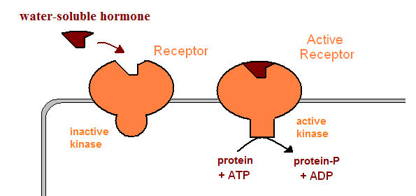
    activation of cytoplasmic kinases by the intracellular domain of the activated receptor
    activation of G-proteins. G-proteins contain three subunits, one of which binds GDP. When a G-protein binds an activated receptor, its GDP-binding subunits undergoes a subtle conformational change, and exchanges its GDP nucleotide by a GTP. The GTP-bound subunit can no longer bind the other subunits, and separates. This isolated GTP-bound subunit may the activate different signal transduction mechanisms:
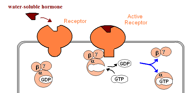
          activation of adenylyl (or guanylyl) cyclases which produce the second messengers cAMP (or cGMP). These second messengers activate specific kinases, which in turn activate (or deactivate) a wide array of enzymes.
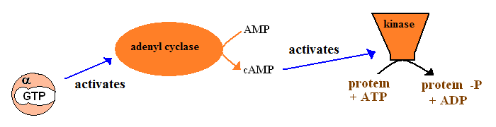
          activation of phospholipase C. This enzyme kydrolyzes a specific phospholipid, yielding two second messengers: inositol triphosphate (IP3) and a diacylglycerol (DAG). DAG activates the (membrane-bound) protein kinase C, and IP3 triggers Ca2+ release from the endoplasmic reticulum.

    The effects triggered by these hormones are transient: e.g. cAMP eventually is hydrolyzed by phosphodiesterase into AMP, terminating its activating effect, phosphatases remove the phosphate group from phosphorylated proteins, etc. Hormones may also be removed from the bloodstream by enzymatic activities present in the liver, or through excretion by the kidneys. Water-soluble hormones are usually the easiet to remove, and most often disappear from the bloodstream in a few hours. On the other hand, lipid-soluble hormones are carried in the plasma by proteins, and remain in the bloodstream for a longer time.
    Hormones which stimulate the secretion of additional hormones are called tropic hormones.

    Pituitary and hypothalamus

    The pituitary (hypophysis) is a gland located under the hypothalamus. It contains two histologically different regions:
  • posterior pituitary (neurohypophysis) is mostly composed of terminal portions of hypothalamic neurons. In the neurohypohysis these neurons come into contact with the blodstream, into which they release two peptide hormones:oxytocin (which stimulates the ejection of milk from the mammary glands and uterine contractions) and vasopressin (also known as antidiuretic hormone- ADH).
  • anterior pituitary (adenohypophysis) contains endocrine cells and communicates with the hypothalamus through a specialized capillary network called hypothalamic-hypophyseal portal system. This network allows the hypophysiotropic hormones released by the hypothalamus to reach the hypophysis before being diluted in the general bloodstream. Six hormones are secreted by the adenohypohysis:
  • FSH - Follicle-stimulating hormone (involved in gametogenesis)
  • LH - Luteinizing hormone (acts on the gonads). FSH and LH are collectively called gonadotropins.
  • TSH (thyrotropin) - thyroid-stimulating hormone
  • ACTH - adrenocorticotropic hormone, also know as corticotropin (acts on the adrenal glands)
  • growth hormone (somatotropin)
  • prolactin - Stimulates the development of the mammary glands and milk production.

  • The hypothalamus controls the adenohypohysis through the secretion of several hypophysiotropic hormones :
  • GnRH - Gonadotropin releasing hormone
  • somatostatin (inhibts growth hormone secretion)
  • dopamine, also know as "prolactin-inhibiting factor" (inhibits prolactin secretion)
  • TRH - thyrotropin-releasing hormone (also stimulates prolactin secretion)
  • GHRH - growth hormone-releasing hormone
  • CRH - corticotropin-releasing hormone

  • Hormone disorders may be due to abnormal amounts of circulating hormones (either hypo- or hyper-secretion) or to abnormal target cell responses (hypo- or hyper-sensitivity). Anomalies of the amount of hormone may be due to malfunctioning of:
  • the secreting gland a (primary hypo- or hyper-secretion)
  • the gland (usually the pituitary) that secretes its tropic hormone( secondary hypo- or hyper-secretion)
  • the gland (usually the hypothalamus) that secretes the regulating hormone of its tropic hormone(tertiary hypo- or hyper-secretion)
  • Anomalies in the magnitude of the target cell response to hormone binding may be due to the amount of receptors present, to inhibition of cellurar mechanisms ellicited by receptor activation or to anomalous maturation of the hormone (several peptide hormones must be activated by hydrolysis after secretion to the bloodstream).

    Thyroid

    The thyroid gland, located in front of the trachea, weighs approximately one ounce. It is composed of several follicles, which contain an outer layer of secreting cells surrounding a solvent-filled cavity where proteins (mainly thyroglobulin) accumulate filled. The thyroid actively absorbs iodide from the bloodstraeam and transfers it (through the action of a peroxidase enzyme system) to specific tyrosine residues present in thyroglobulin. Eacy tyrosine may receive one or two iodine atoms (yielding either monoiodotyrosine -MIT_ or diiodotyrosine - DIT). Iodine-containing hormones produced in the thyroid are built from these iodinated tyrosine residues: thyroxine (T4) is built from two DIT molecules, and triiodothyronine (T3) is built from one DIT and one MIT. Both T4 and T3 are lipid-soluble hormones, and are carried in the bloodstream by specific proteins (like transthyrretin). T4 is much more abundant than T3, but only T3 is active: T4 must be deiodinated to the active form (T3) by the target cell.
    Iodine-containing thyrois hormones induce the synthesis or respiratory enzymes, Na+-K+ ATPase, etc. induzem a síntese de enzimas respiratórias, da bomba de Na+-K+, etc., which causes a general increase of the metabolic rate and heat production. Their effects on the body also include appetite increase, decrease of adipose tissue, increased breaathing rate and urea excretion, etc.
    Whenever the concentrations of T3 and T4 in the bloodstream are too low, TSH is secreted by the pituitary gland. TSH stimulates the thyroid to increase its protein production and size. Therefore, in the absence of iodine the size of thyroid gland increases considerably due to its continuous stimulation by TSH.

    Hormone regulation of kydney function

    In the kydney's Bowman capsules, blood plasma is continuously filtered. Apart from the absence of proteins, the composition of the filtrate inside the capsule is identical to that of the plasma outside the capsule. The collected filtrate flows through the kydney tubules, where several molecules are either reabsorbed into the bloodstream, or secreted into the filtrate. The composition of the resulting outflow (urine) is therefore quite different from that of the original filtrate.

    Further reading:

    cover
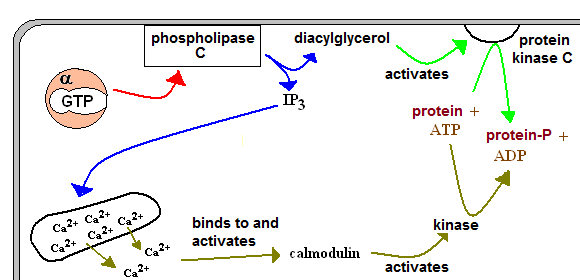
Human Physiology: The Mechanisms of Body function

    A good text for undergraduate students.

    coverTextbook of Medical Physiology

    A classic on Human Physiology. Excellent and detailed, it is invaluable for Nursing and Medicine students.

    visitas desde 4 de Janeiro de 2002